X-Ray Interpretation
Gout is caused by the deposition of monosodium urate crystals. About 50% of patients manifest radiographic bony changes, usually 6-8 years after the initial attack. These changes depend on the sites where the urate crystals are deposited. The following are features of chronic tophaceous gout.
ASYMMETRICAL POLYARTICULAR DISTRIBUTION
The feet and hands are commonly involved. Joint involvement can occur in a very asymmetric (random) polyarticular pattern.
TOPHI
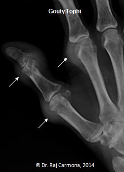
Tophi are soft tissue masses created by the deposition of urate crystals. Urate is not inherently radio-opaque. The varying densities seen on radiographs is due to calcium precipitation with the urate crystals. Tophi are typically located in the peri-articular area along the extensor surface, but maybe intra-articular or not associated with the joint at all. The following radiographic shows tophi at the 1st and 2nd MCPs and the IP joint of the thumb.
PUNCHED-OUT EROSIONS
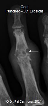
Erosions can occur in a very sporadic asymmetric distribution. Borders can be sclerotic because of the indolence of the process, creating a punched-out or “mouse bitten” appearance. The following radiograph shows a typical erosion at the PIP (right side of radiograph).
INTRA-OSSEOUS LESIONS
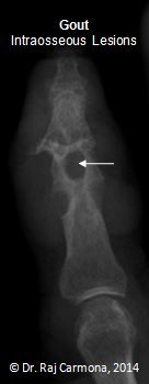
Urate crystals are deposited within the bone producing an intraosseous tophus with or without calcification. This is seen as a lytic lesion and can be expansive.
SCLEROTIC OVERHANGING EDGES
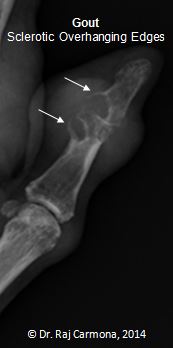
As the erosion develops, the edges of the cortex can be remodelled in an outward fashion, creating an overhanging edge. The following radiograph vividly demonstrates this at the IP joint of the thumb.
Additionally, because of the chronicity of the process, bone production leads to sclerotic borders, overhanging edges, bony enlargement, and irregular spicules at the sites of tendon and ligament attachments.
PRESERVED JOINT SPACE
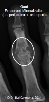
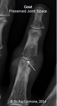
Joint spaces are preserved until late. Even if the joint is partially destroyed (usually on the dorsal aspect), preservation of another area of the joint (usually flexor aspect) creates the radiographic impression that the joint space is preserved. This is demonstrated at the PIP joint in the following radiograph.
NO PERIARTICULAR OSTEOPENIA
In contrast to rheumatoid arthritis, mineralization is maintained and there is no predominant periarticular osteopenia. Despite multiple erosions at the PIP joint in the following radiograph, there is no osteopenia.
